Check out the small ‘pizza piece’ down to the nerve cell.
Lauren Leffer
|
Released May 9, 2024 2:00 PM EDT
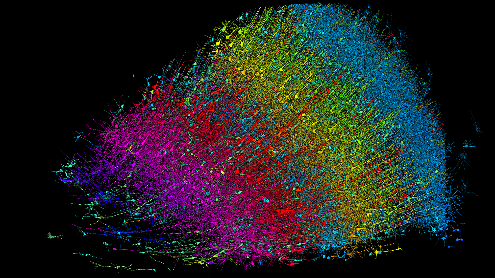
A cubic millimeter is, by all accounts, small. It’s hardly visible– a speck or fleck or crumb. Look carefully sufficient and you can discover a whole world inside a particle of product. A group of neuroscientists and engineers, assisted by artificial intelligence tools, have actually charted a cubic millimeter volume of the human brain at nanoscale resolution, tracing every nerve cell, synapse, capillary, and supporting cell within the piece and rebuilding a 3D design of the tissue. It represents simply one-millionth of the overall brain volume, it is the most comprehensive map of a piece of human brain matter ever developed. It might stimulate a wave of clinical discovery about neurological conditions, brain structure, and the origins of our habits.
“In one regard, our information set is small,” Jeff Licthmanco-senior research study scientist and a neuroscientist and teacher of molecular and cellular biology at Harvard University, informs PopSci“But it does not feel little due to the fact that when you get in it, you see it’s like an enormous forest. It’s a really small forest, however it’s a really, extremely, extremely complex forest,” he includes.
All that intricacy is on display screen in a research study recording the building of this little bit of detailed brain map or “connectome,” released May 9 in the journal ScienceThe very first connectome was of a nematode brain, finished in 1986Ever since, neuroscientists have actually continued to outline out significantly big and complex brains– consisting of those of fruit flies, maggots, a tadpole, and an earthworm. Human brains present a distinct mapping difficulty in their complexity and inaccessibility. The brand-new, partial human connectome is readily available online for anybody to check out.
“Not just is this an outstanding technological task, this is a tool and a resource that is actually targeted at showing the world and getting all of this clinical details out there,” Tim Moscaa neuroscientist at Thomas Jefferson University who was uninvolved in the brand-new work, informs PopSci“This group has actually done a remarkable task creating all of the brand-new tools and the pipelines to make this offered to anybody who wishes to take a look at it, wishes to think of it, wishes to utilize this in their research study.”
Dishing out brain pizza
The research study sample was gathered over a years earlier from a confidential client going through epilepsy surgical treatment. The cosmetic surgeon got rid of a little piece of the temporal lobe to gain access to and deal with a hidden sore, rapidly maintained the tissue, and later on shared it with researchers. The overall volume of the piece is about 1 cubic millimeter, it is not cube formed. Rather, “it’s like a thick piece of pizza– however it’s not that thick,” states Lichtman. This blunt, triangular portion, longer than it is broad, made it possible for the scientists to record a little all 6 layers of the 3mm thick cortex.
The primary step to mapping the brain pizza was to slice it into 5,019 specific sample (each 30 nanometers thin) installed on tape utilizing a specifically developed device that cuts with a diamond knife. From there, the scientists invested a complete year thoroughly imaging each piece by means of electron microscopic lense. They digitally lined up and sewed together the pieces and utilized numerous maker knowing tools to fill out the 3-D kind and label and color each part.
The sector’s nerve cell density is 16,000 nerve cells per cubic millimeter– about one-third lower than a previous density quote of the exact same brain area and 10 times less thick than the matching area of a mouse’s brain, per the research study. Glial cells, the connective glue that keeps brain tissue together, surpass nerve cells in the piece by a 2 to one ratio.
Neural explorers
The physical size of the brain piece might be tiny, however the level of information implies the information caught by the mapping effort is huge. The rebuilt section is 1.4 petabytes in digital size, or 1,400 terabytes (comparable to the storage capability of about 2,800 typical laptop computers). Within that, there is lots to possibly find: specific neural circuits, formerly unnoticed cellular ratios and shapes, the makeup of each cortical layer, and more.
“It’s like being an explorer that arrive on a brand-new island,” states Lichtman. “You keep browsing and you’re simply going to keep discovering brand-new things.”
Currently, Lichtman and his numerous co-researchers have actually made some fascinating observations. In the middle of the ~ 150 million synapses they mapped, they discovered an uncommon kind of especially strong connection. In the huge bulk (96.5%) of cases, axons– the outbound transmission line of nerve cells– formed one connection with a target cell. Some (about 3%) made 2 connections. Less than.01% created more than 4 synapses, consisting of some axons and target cells that were linked at over 50 points.
“We’ve constantly had a theory that there would be very connections, if you will, among particular cells,” states Mosca. “But it’s something we’ve never ever had the resolution to show … Now we understand that it exists and we can pursue the concern of what it does.” Lichtman’s present hypothesis is that these extra-fortified connections are the sort of hyper-fast paths that allow “automated usage of the brain” for reputable, found out actions.
Another brand-new observation: lots of dendrites (the branching extensions of nerve cells that normally get inputs) appear to mirror each other– pointing symmetrically in among simply 2 directional plans out of limitless three-dimensional possibilities. “We ‘d never ever seen anything like that [before],” states Lichtman. “Why are they doing that? We do not understand … [it is] a total secret.”
The researchers even more discovered a brand-new kind of inexplicable structure that they’ve called an “axon whorl,” where long axon cable televisions appear to tangle around themselves. It wasn’t every nerve cell, some axons consisted of numerous knots, states Viren Jainco-senior research study author and a senior personnel researcher at Google where he leads the business’s Connectomics research study group. Once again, the function and reason for these whorls is unidentified. “We were not anticipating to discover such a structure. It’s extremely strange … like a huge assortment of electrical wiring that sort of contravenes the function of a wire to start with, which is to go locations and contact other things.”
These 3 findings are most likely simply the pointer of the iceberg. “The information set is so big that a person human being or laboratory group can not explore it [all]however a lot of people can,” Lichtman states. Due to the fact that of the open nature of the task, more than 200 documents have actually mentioned the brain restoration because it was very first launched as a pre-print, Jain explains.
In addition to being a big, essential advance in science, discoveries arising from this partial connectome might ultimately assist us much better comprehend and deal with brain illness. “The capability to determine neural circuitry of human brains in such information opens amazing chances for advancing human health,” states Andrew Leifera physicist and neuroscientist at Princeton University who wasn’t associated with the task. “One might picture comparing various brains to comprehend how brain electrical wiring modifications when a healthy brain struggles with an illness or falls under dysfunction,” he includes.
Pressing into future frontiers
Though there is lots to be found, there are likewise limitations. The automated device finding out techniques which were essential to allowing such a massive venture bring a margin of mistake that needs human oversight to remedy. Modifying will be a continuous task, and is a neighborhood science effort anybody who wishes to can use to take part in
The sample is likewise just one little piece of someone’s brain. There is much that can’t yet be presumed about human brains normally or other brain areas beyond the temporal lobe based upon this single piece without more samples and maps for contrast, keeps in mind Lichtman.
And, maybe most seriously, the brain sector originated from somebody going through surgical treatment for epilepsy– it might not represent a “typical” brain and there’s no other way to understand for sure unless and till we have more bits to evaluate, state Jain and Lichtman. “But we are preparing lots of follow-ups to this,” Jain includes.
The group has aspirations to build several partial connectomes representing extra human brain samples. They are likewise dealing with zebrafish connectome, and are preparing to take on progressively big sections of the mouse brain. Mammalian brains share lots of resemblances, so a total mouse connectome might provide brand-new insights into our own brain in addition to the advancement of brains throughout animals, Lichtman states.
At the minute, with presently readily available innovations (and the ethical ramifications), a total connectome of the human brian is “a bridge too far,” states Lichtman. “Literally, we’re a million times far from that,” states Jain. Through this research study, the researchers have actually taken an early (if little) action in that instructions, and even the tiniest peephole can be a website into an entire universe of understanding.
“I would enjoy individuals to consider this the very same method they consider the Hubble or James Webb telescope,” states Lichtman. “We’re peering into an unidentified domain, and one that is a lot more pertinent to us than far-off deep space. It’s this inner area that each people have on our shoulders that we utilize, however understand practically absolutely nothing about.”